
Extracorporeal Shock Wave Therapy
EXTRACORPOREAL SHOCK WAVE THERAPY
QUICK - SAFE - SUCCESSFUL
ESWT offers PAIN RELIEF & RESTORATION of MOBILITY in:
- TENDON Injuries
- LIGAMENT Injuries
- MUSCLE Pain
- Delayed BONE Healing / Stress Fractures
- JOINT disease such as Osteoarthritis
NEW DISCOVERY :
Is There A Role For ESWT In Wound Care?
VOLUME: 19 PUBLICATION DATE: Jul 01 2006
Issue Number:
7
Author(s):
By John S. Steinberg, DPM, Lt. Col. Alexander Stojadinovic, MD, LCDR Eric Elster, MD, Lt. Col.(P) George Peoples, MD, and Chris E. Attinger, MD
Over the past several years, there has been a developing body of knowledge regarding the clinical applications of extracorporeal shockwave therapy (ESWT). The latest area of clinical investigation for this technology is in the arena of wound healing. Researchers are now studying ESWT as a new approach to wound healing with a particular emphasis on complex soft tissue wounds with and without underlying bone disruption. Hopefully, this article will serve as an introduction to this new topic and we hope the evidence-based data will soon follow as the ongoing clinical trials progress.
Preliminary clinical trials show that the core technology of unfocused ESWT (Tissue Regeneration Technologies) significantly enhances and accelerates the healing of complex soft tissue wounds in comparison to standard methods of treatment. Experience to date with ESWT indicates it is uniquely suited to the challenge of efficiently and effectively treating a variety of complex wound types, including military combat wounds. If proven in the proposed definitive field testing, such a system would offer new treatment options for some of our most challenging non-healing wound types.
Clinicians have used ESWT successfully to disintegrate kidney stones since 1981. The high efficacy and few adverse effects associated with this treatment made it the standard of care worldwide. Then shockwave therapy became a standard treatment for various common orthopedic conditions such as plantar fasciitis, tendinosis calcarea of the shoulder and tennis elbow.1 Further research has demonstrated shockwave therapy's effectiveness in treating nonunion fractures in long bones and aseptic bone necrosis in humans.2-4 Unlike the treatment of kidney stones, the fundamental therapeutic objective of orthopedic shockwave application is not to destroy tissue but to stimulate tissue regeneration. 5,6
Researchers have shown that treatment with ESWT increases the release of critical wound growth factors and promotes vessel in-growth. The resulting improved circulation within the wound can have certain benefits in a variety of wound etiologies. Animal studies indicate that local delivery of shockwave therapy stimulates early expression of angiogenesis-related growth factors, including endothelial nitric oxide synthase, vessel endothelial growth factor and proliferating cell nuclear antigen. This results in new vessel in-growth that improves blood supply, increases cell proliferation and accelerates tissue regeneration and healing.1,7-9
Ludwig, et. al., described the beneficial effect of ESWT on wound healing in 1990. 10 During the course of treating nonunion or delayed osseous union of bone fractures, researchers discovered that both the disrupted bone as well as overlying soft tissue wounds healed rapidly. 3 Subsequently, an extensive body of peer-reviewed literature emerged, demonstrating that one could apply ESWT safely and with minimal risk to treat patients with a variety of complex surgical problems, including nonunion of long bone fractures with or without osteomyelitis, aseptic femoral head necrosis, pseudoarthroses and osteochondritis dissecans. 10-15 The observed antibacterial effects of extracorporeal shockwaves are highly relevant to the increased risk of infection common to non-healing wounds.
Understanding The Different Mechanisms Of ESWT
While the ESWT technology is quite different from current standards of soft tissue wound care, it is conceptually similar in its potential wound benefits. Extracorporeal shockwave therapy appears to increase local wound vascular density and blood flow as well as the rate of granulation tissue production while reducing bacterial counts. This would clearly accelerate healing time and promote the eradication of preexisting bacterial overgrowth. Extracorporeal shockwave therapy has shown potential for spontaneous epithelialization with a reduced need for secondary suturing, skin graft or flap closure.
Extracorporeal shockwave therapy generates an acoustic pressure wave that penetrates human tissue through a liquid medium. One can focus the energy wave to a point or distribute it over a broad therapeutic front. Initially, researchers believed ESWT mechanically induced micro-sized fractures in bone tissue. However, it is now widely accepted that ESWT induces a reproducible biological response in target and surrounding tissue, releasing wound growth factors locally, producing an antibacterial effect on biofilms (clusters of bacteria with a protective coating). Accordingly, ESWT facilitates vascular in-growth, re-epithelialization and complete wound closure.
Current versions of ESWT in clinical use are based on the technology of an electrohydraulic shockwave. To achieve this, one would position a spark plug with two opposing electrodes under water in an ellipsoid reflector, releasing an electrical discharge in the focal point F1 (between opposing electrodes). The opposing electrodes are connected to a capacitor, which one charges to the maximum voltage and then abruptly discharges. The underwater discharge causes the explosive formation of a plasma channel, which results in evaporation of the water surrounding the opposing electrodes.
The primary spherical shockwave that is released at F1 expands in the surrounding water and the walls of the ellipsoid reflector reflect the shockwave. Due to the geometric properties of an ellipsoid reflector, all shockwaves generated at the opposing electrodes (F1) are reflected to a small area at a fixed distance away (known as the focal point or F2) from the opposing electrodes. Therefore, during an ESWT procedure, one should carefully align the device shock head and the patient, ensuring the target area to be treated is located at F2 where F2 is typically the point of maximum pressure.
In considering the application of ESWT to wound care, the technology must be adapted into a wide focus treatment area rather than the highly focused devices currently in use. To achieve shockwaves that are "roughly plane" to the treatment area, the reflector utilized in the wound treatment device applicator is made of a generalized parabolic reflector.
Placing the tips of the electrode (F1) in an exact parabolic reflector will result in a plane wave which emits after the reflection of the primary spherical wave. The focal point (F2) of these plane waves is, by definition, "unfocused" or "defocused," meaning a focal point (F2) no longer exists. Therefore, the shockwave characteristics for the parabolic reflector could be defined as plane waves or, in other words, parallel rays. By using the generalized parabolic reflector, the plane waves are bent slightly toward the central acoustic axis. Therefore, the waves are unfocused, nearly parallel and a focal point (F2) does not exist. The energy density realized by the generalized parabolic reflector is higher than with an exact parabolic reflector and the acoustic field stimulates a large area.
How Clinicians Would Use ESWT To Treat Wounds
While all of this sounds quite complex, the actual use of the ESWT device in the wound patient is quite simple. One would prepare the patient with appropriate sharp debridement and local wound care. Then place the ESWT applicator head over the wound, utilizing ultrasonic gel and plastic draping to prevent any cross-contamination of the device.
The current study protocol utilizes 100 shocks per 1 cm2 of wound so the treatment lasts just one to two minutes. The most sensation the patient may notice is the sound of the ESWT machine as it generates the shockwaves and bright flashes of light that correspond to those waves.
Currently, several shockwave devices are FDA approved to treat kidney and ureteral stones, chronic heel pain (plantar fasciitis) and tennis elbow (lateral epicondylitis). The devices have proven useful in treating Achilles and patellar tendonitis as well as challenging and difficult to treat delayed osseous union or nonunion fractures as well as avascular necrosis. Under European CE medical device approval, ESWT is routinely used by physicians to treat chronic non-healing soft tissue wounds, ischemic heart disease, arthritis, burns and periodontal disease successfully.
What Does The Research Indicate?
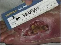
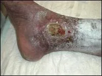
Clinical research conducted by Schaden at the Meidling Trauma Center in Vienna, Austria and at the Berlin Center for Extracorporeal Shock Wave Therapy suggest that ESWT is very effective in treating acute and chronic skin lesions such as venous and arterial ulcers, decubitis ulcers, post-traumatic wounds and burns.15 The study was presented in May at the 9th International Society for Musculoskeletal Shockwave Therapy in Rio de Janiero. The study involved 200 patients including 65 with post-traumatic lesions, 25 venous ulcers, 28 arterial ulcers, 13 decubitus ulcers and five burn injuries.15
Researchers utilized a DermaGold™, MultiWave™ MTS 180 (Tissue Regeneration Technologies). Since surface defects are often involved, researchers modified the shockwave head so the shockwave would no longer be focused in a small plane to the treatment area. Clinicians used low energy flux densities and, depending on the size of the defect, the number of pulses varied between a few hundred to several thousand. Researchers administered no antibiotics (unless the patient was already taking them), anesthesia or debridements in the study.
Between September 2004 and January 2005, researchers used ESWT to treat 104 non-healing soft tissue wounds in 102 patients (50 male and 52 female patients with a median age of 61 years) with ESWT. They received 100 to 1,000 shocks/cm2 at 0.1 mJ/mm2, according to the size of the wound. The researchers administered the treatments weekly or every other week based on the rate of regeneration over one to six treatments (with a mean of three treatments). The rate of epithelialization was carefully documented. The mean treatment time was three minutes per session.
The pre-ESWT wound dressing therapy was not modified for the purpose of this study and continued after patients were treated with ESWT. For clean wounds/ ulcers, researchers employed wet-to-wet dressings with Tender-Wet®. Wounds with heavy secretions received Seasorb® or Comfil®, and the researchers used Aquacel Ag® for wounds with necrotic tissue.
Despite the fact that the wounds had bacterial contamination, no patients received antibiotic therapy during ESWT. No anesthesia was necessary in any of the patients studied as the shockwave was defocused and delivered over a broad treatment front. Researchers administered all ESWT on an outpatient basis and patients tolerated it well without any adverse side effects. Nine patients did not complete treatment and constituted study dropouts. (See "A Closer Look At Healing Rates Of Wounds Treated With ESWT" above.)
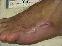
During a three- to 12-week period of monitoring, 81 percent of wounds healed completely with 12 percent demonstrating incomplete healing but greater than 50 percent epithelialization.
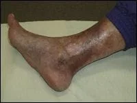
Throughout the course of this pilot study, none of the wounds deteriorated with ESWT. Interestingly, obviously colonized wounds at the outset of treatment improved without antibiotics after the first treatment session, presumably due to the bactericidal effect of shockwave therapy. No wound infection developed in this patient cohort. Surprisingly, even chronic venous stasis and arterial insufficiency ulcers demonstrated rapid wound healing with ESWT as 19 (53 percent) of 36 patients healed completely within six to 12 weeks while 10 (28 percent) patients had epithelialized over 50 percent of the treated wound surface area. No patient in this study experienced any deterioration of the treated wound.
Where Is The Current Research On ESWT Headed?
Based on these encouraging results and other studies throughout the world, an FDA-sponsored diabetic foot ulcer study began in the United States earlier this year. Chris Attinger, MD, and John Steinberg, DPM, of the Limb Center at Georgetown University, have begun enrolling patients into this randomized, controlled trial for diabetic foot wounds using the wide focused ESWT technology. Several other sites will begin enrolling patients in this study as this article is published.
Very promising research involving similar ESWT technology is currently proposed for military combat wounds. Eric Elster, MD, George Peoples, MD, and Alexander Stojadinovic, MD, are leading the way in a pivotal randomized trial that will evaluate the efficacy of the technology in the management of acute traumatic wounds of the extremity with an emphasis on combat wounds.
Clearly, the possible utility of this technology is just in the process of emerging into wound care. The ongoing and future clinical trials, which employ randomization and specific wound types, should yield definitive clinical evidence on whether ESWT can play a role in healing the problem wound. We look forward to seeing the results of the clinical trials for proper assessment of the position that this may take within the current treatment protocols.